12Lead ECG Placement Guide with Illustrations
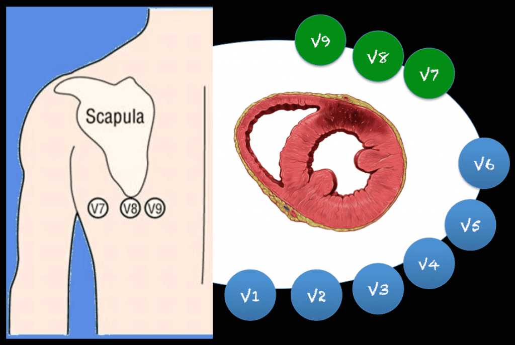
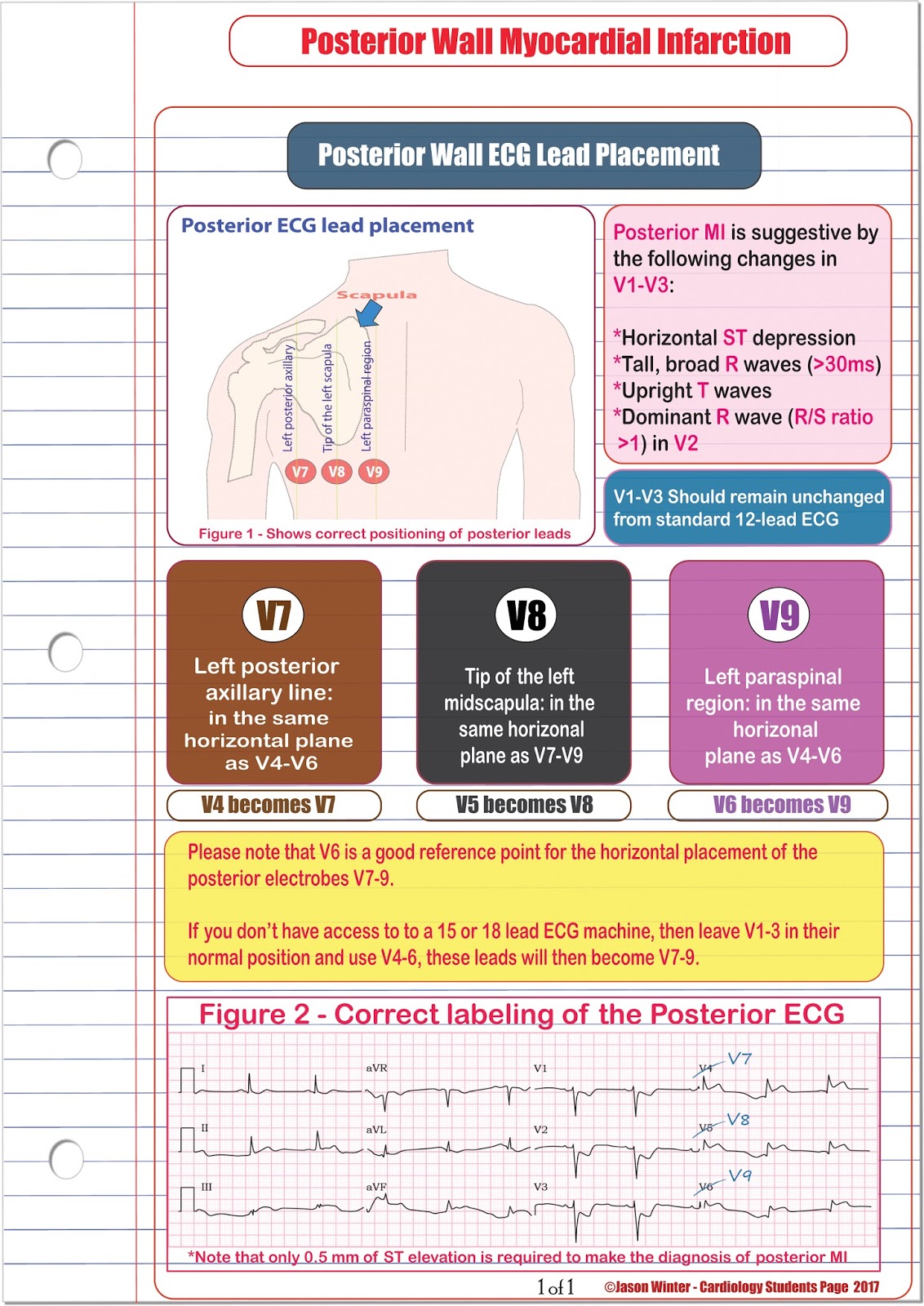
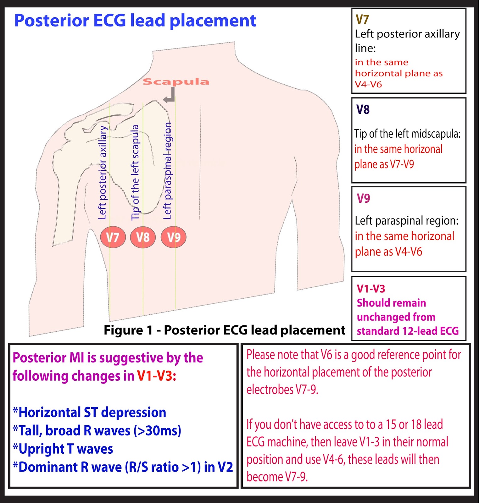
In uncertain cases, a posterior ECG can be obtained by placing posterior leads V7, V8, and V9 below the patients left scapula along the same horizontal plane as V6. At least 0.5mm of ST elevation in one lead indicates posterior STEMI. Posterior ECG lead placement. Case continued: The emergency provider recognized this as highly suspicious for.
ECG Lead positioning • LITFL • ECG Library Basics
Posterior extension of an inferior or lateral infarct implies a much larger area of myocardial damage, with an increased risk of left ventricular dysfunction and death. Isolated posterior infarction is an indication for emergent coronary reperfusion.

Download figure Ekg placement, Nursing information, Medical drawings
What are ECG criteria for posterior MI on the standard 12-lead ECG? 1 R/S wave ratio >1.0 in lead V2 Co-existing acute, inferior, and/or lateral MI Limited to leads V1 - V3: ST-segment depression (horizontal moreso than downsloping or upsloping) Prominent R wave Prominent, upright T wave

ECG Educator Blog Posterior ECG Lead Placement
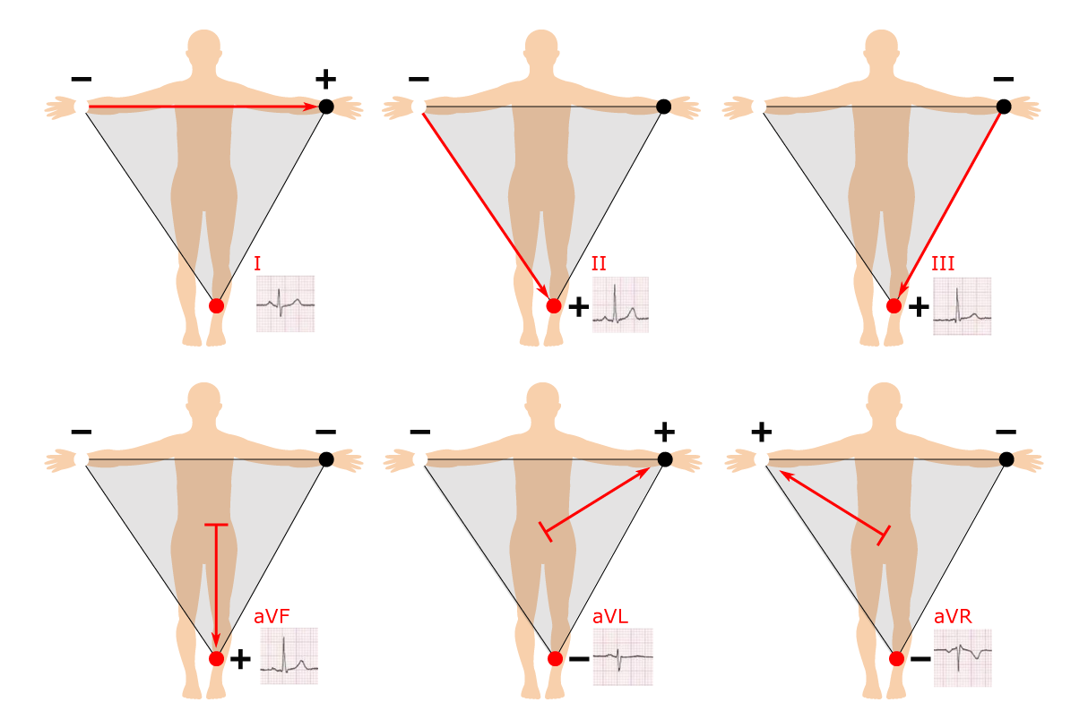
An ECG lead is a graphical description of the electrical activity of the heart and it is created by analyzing several electrodes. In other words, each ECG lead is computed by analyzing the electrical currents detected by several electrodes.
Five ECG Patterns You Must Know R.E.B.E.L. EM Emergency Medicine Blog
An American Heart Association Scientific statement reports that recordings from right-sided and posterior ECG leads increases the diagnostic sensitivity for STEMI. 3 However, additional leads required electrode repositioning and a change in patient positioning to obtain V3R-V6R and V7-V9. For this reason and others, they are not routinely done.
ECG Educator Blog Posterior ECG Lead Placement
ECG lead placement in a posterior body position is not a unique concept. Classically posterior leads V 7 to V 9 , have been used as an extension of precordial leads V 1 to V 6 in the detection of posterior myocardial infarction ( 21 ).

v7 v8 v9 lead placement checkmatepaintingparisfrance
12 Lead ECG Placement Guide. The correct positioning of leads is essential to taking an accurate 12 lead resting ECG and incorrect placement of leads can lead to a false diagnosis of infarction or negative changes on the ECG. This guide explains the common position for each of the 10 leads on a 12 lead resting ECG. Electrode.

Posterior Myocardial Infarction • LITFL • ECG Library Diagnosis
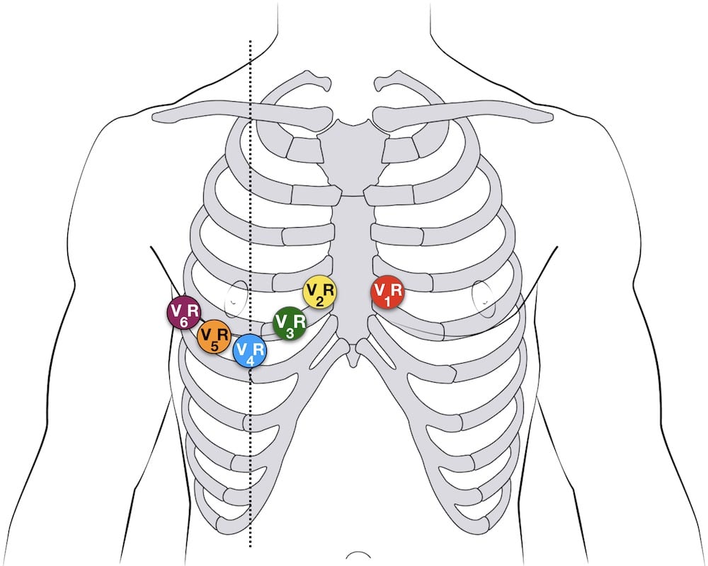
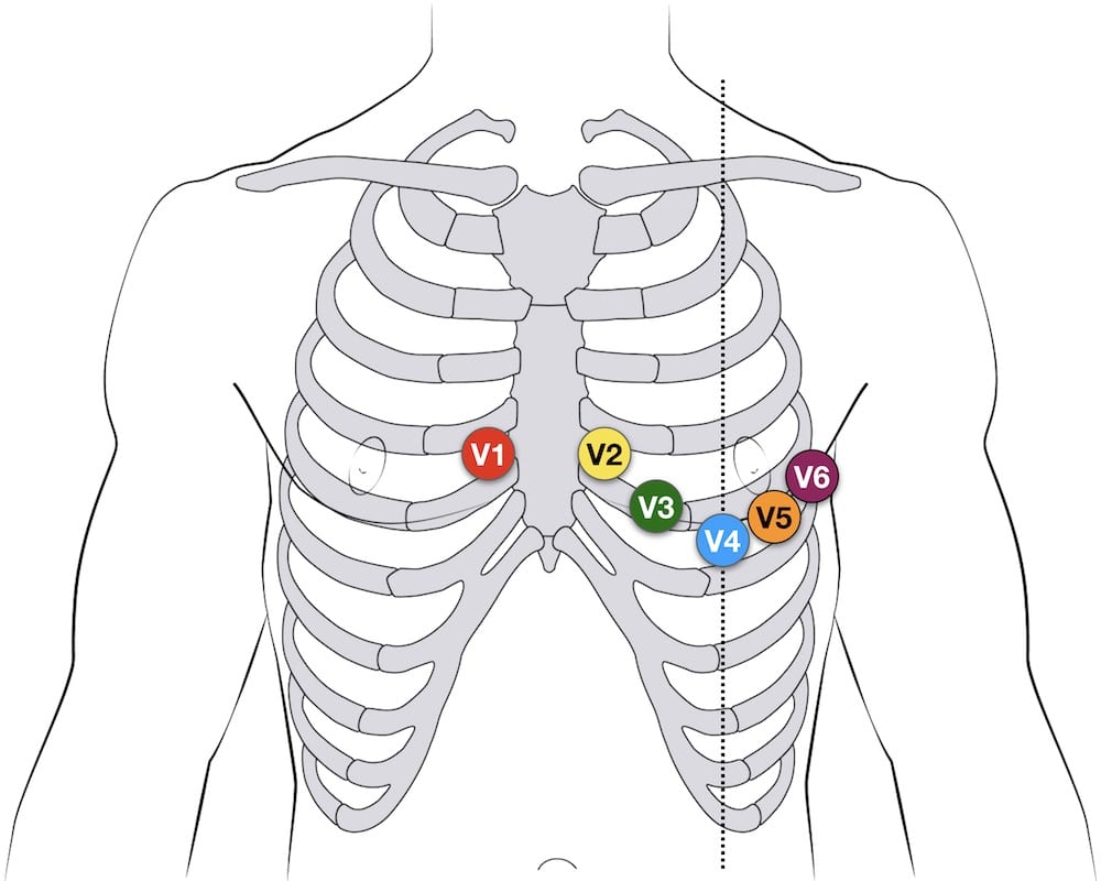
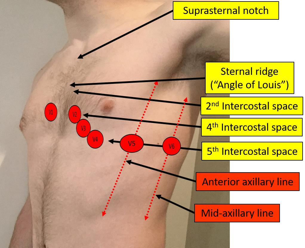
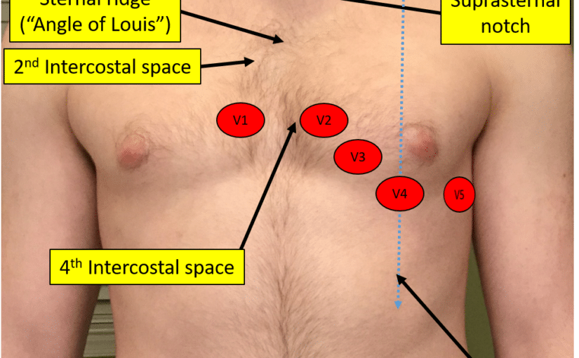
The easiest way to correctly place the electrodes is in the following order: V1 - 4th intercostal space, just to the right of the sternum. V2 - 4th intercostal space, just to the left of the sternum. V4 - On the mid clavicular line & 5th intercostal space. V6 - On the mid axillary line, horizontal with V4. V5 - Between V6 & V4.

Posterior Ecg Lead Placement / ECG introduction at University of Texas Health Science
The initial 12-lead ECG ( Figure 1A) was obtained with limb leads placed on the torso position and reported as "ST elevation and possible acute inferior wall myocardial infarction (MI)". Repeat ECG with limb leads placed in standard, distal limb positions showed resolution of "ST elevation" in the inferior leads ( Figure 1B ). Figure 1A
ECG Lead positioning • LITFL • ECG Library Basics
Background For decades, I noticed a significant inconsistency in the way electrocardiograms are performed. I asked nurses, EKG technicians, medical assistants, and even cardiology fellows where ECG leads/electrodes should be placed on the patient's body.

ECG placement & misLEADing ECG’s EMbeds.co.uk
To detect posterior STEMI associated with occlusion of the circumflex artery or dominant right coronary artery, obtain a posterior ECG. 2-3 [Level A Recommendation] When a 15-lead &/or 18-lead ECG machine is not available, manipulation of the leads from a standard 12-lead ECG machine allow additional areas of the heart to be imaged.4-5
ECG Educator Blog Posterior ECG Lead Placement
ECG lead placement in a posterior body position is not a unique concept. Classically posterior leads V 7 to V 9 , have been used as an extension of precordial leads V 1 to V 6 in the detection of posterior myocardial infarction ( 21 ).
Proper Electrocardiogram (ECG/EKG) Lead Placement ECGEDU
As posterior leads, when an eletrocardiogram with right-side leads has been made, to avoid confusion, you must write the word Right in the electrocardiogram header, and must write also a letter R after the name of replaced precordial leads (V3-V6). This clarify that it is a right-side electrocardiogram. previous | next If you Like it. Share it.
Proper Electrocardiogram (ECG/EKG) Lead Placement
Thus, the 12-lead ECG does not display ST segment elevations during posterior (posterolateral) transmural ischemia. On the other hand, leads V1-V3 (occasionally V4) may detect the injury currents and present them as ST segment depressions. These depressions are reciprocal ST segment depressions, meaning that they mirror the ST segment elevations.
Lead Placement for Posterior ECG Resus Review
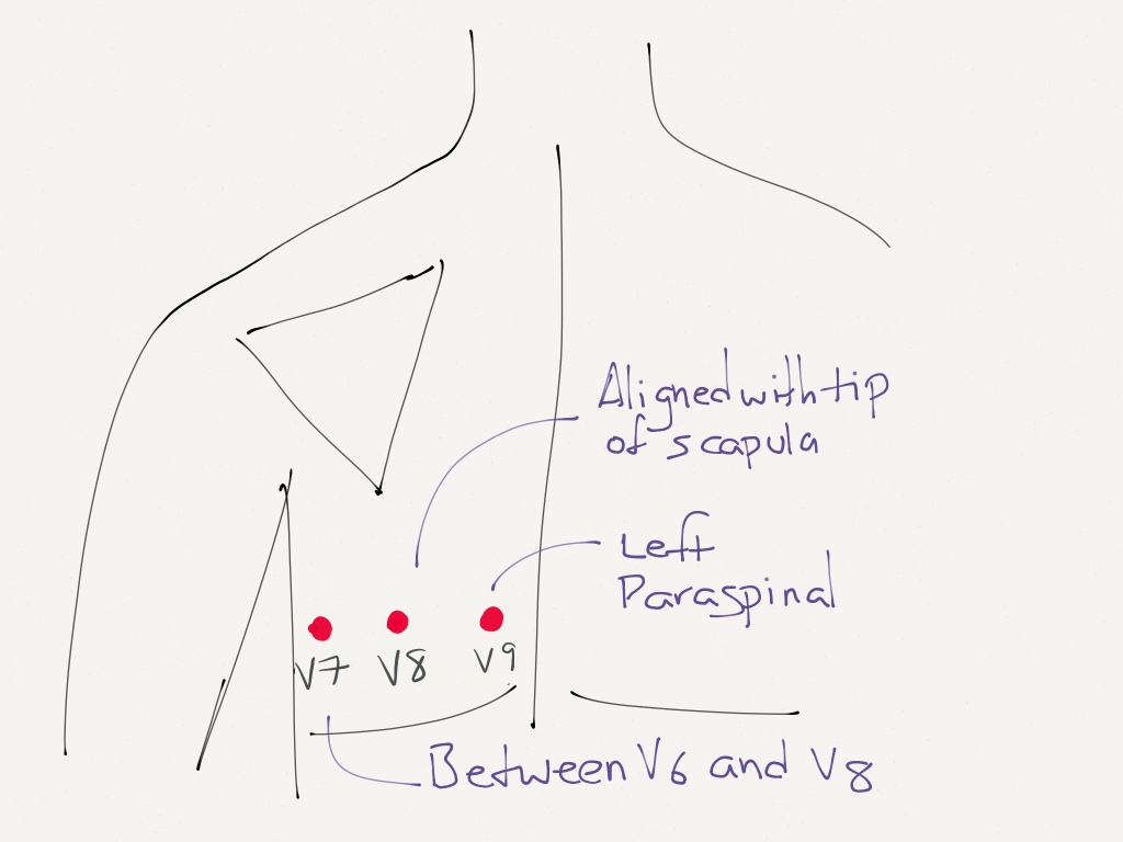
The addition of posterior leads V 7 to V 9 significantly increases the ability to detect posterior injury patterns compared with the standard 12-lead ECG. 5, 7 Lead V 7 should be placed at the level of lead V 6 at the posterior axillary line, lead V 8 on the left side of the back at the tip of the scapula, and lead V 9 halfway between lead V 8 and the left paraspinal muscles.

Telemetry Technician Course EKG Lead Placement (class 3)
5-electrode system Uses 5 electrodes (RA, RL, LA, LL and Chest) Monitor displays the bipolar leads (I, II and III) AND a single unipolar lead (depending on position of the brown chest lead (positions V1-6)) 5-lead electrode placement 12-lead ECG 10 electrodes required to produce 12-lead ECG